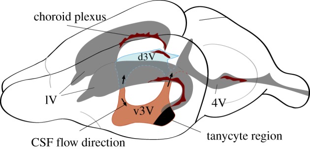
Figure 2.

The four CSF-filled ventricles in the adult mouse brain. Highlighted in colour are the dorsal (d3V, blue) and ventral (v3v, light brown) parts of the third ventricle. The two lateral ventricles feed via a canal into the mid-plane located 3V. At the site of junction, CSF flows either dorsally (up arrow) into the d3V or ventrally (down arrow) into the v3v. At their back end, d3V and v3v connect via the aqueduct into 4V. Most of the lining of the ventricles consists of ependymal cells (E1 cells). The dark-shaded area in v3v consists primarily of α- and β-tanycytes. Dark brown features represent the secretory epithelium of the choroid plexi that releases CSF and secretes a great variety of small and macromolecular solutes. Dorsal is on the top and anterior to the left.
