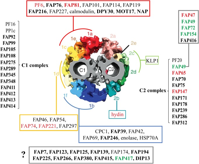
Figure 1.
Summary of CA proteins and their predicted locations in the C1 and C2 microtubules. Diagram of cross-section of the Chlamydomonas reinhardtii CA (modified from [1]) showing predicted locations of CA proteins including novel candidate or confirmed CA proteins identified by Zhao et al. [2] (bold font); FAP76 and FAP216 were localized by Fu et al. [3]. ‘1a’–‘1f’ and ‘2a’–‘2e’ indicate projections C1a to C1f and C2a to C2e, respectively. ASH-domain proteins are in red font; PAS-domain proteins are in green font. Some proteins are predicted to be associated with either the C1 or C2 microtubule, but their specific locations are not yet determined; others (red, green, turquoise, dark blue and yellow boxes) are predicted to be associated with specific projections, pairs of projections or a supercomplex consisting of the C1a, C1e and C1c projections. The FAP47 complex (box, upper right) is likely to be associated with C2 based on solubility properties of FAP49. The question mark indicates proteins whose locations in the CA are not yet known. Modified from Zhao et al. [2]. (Online version in colour.)

