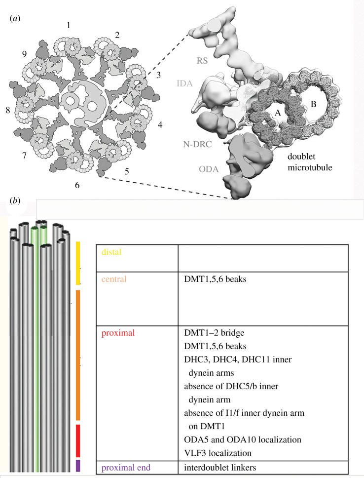
Figure 1.
Diagrams of the Chlamydomonas cilium. (a) Cross-sectional view of the cilium with one doublet microtubule enlarged to show a radial spoke (RS), an inner dynein arm (IDA), the N-DRC and an outer dynein arms (ODAs) as well as the A and B microtubules of the doublet microtubule. The DMTs are labelled 1–9. On doublet microtubule 1 (DMT1), there is no outer arm dynein. The central pair microtubules reside in the middle. (b) A longitudinal view to see the four regions and the unique features of these regions along the length the Chlamydomonas axoneme.

