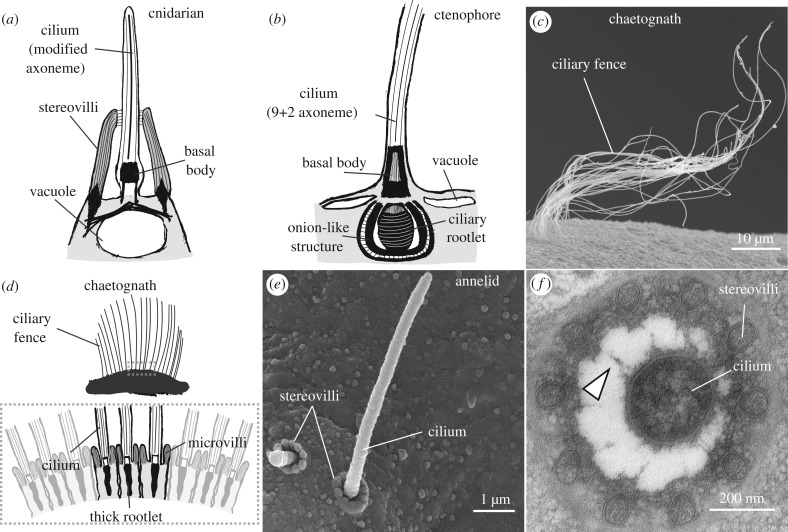
Figure 3.
Structural diversity of vibration receptors. (a) Sketch of a cnidarian vibration receptor. The cilium has a modified axoneme, stereovilli and a vacuole at the base. (b) Sketch of a putative ctenophore vibration receptor. Note the absence of microvilli and the onion-like structure at the base. (c) SEM micrograph of a chaetognath ciliary fence. (d) (Top) Sketch of a ciliary fence in a chaetognath. Dashed rectangle highlights the region expanded below. (Bottom) Sketch of the ciliary fence receptors showing their collared cilia and their tight arrangement along the fence. (e) SEM micrograph of a collar receptor in the annelid P. dumerilii. (f) TEM micrograph of a collar receptor. Arrowhead points to fibres connecting the cilium and the stereovilli. Sketches in a, b and d are based on images/schematics in [112,116], [118–120] and [121,122]. Image in f is taken from [123].

