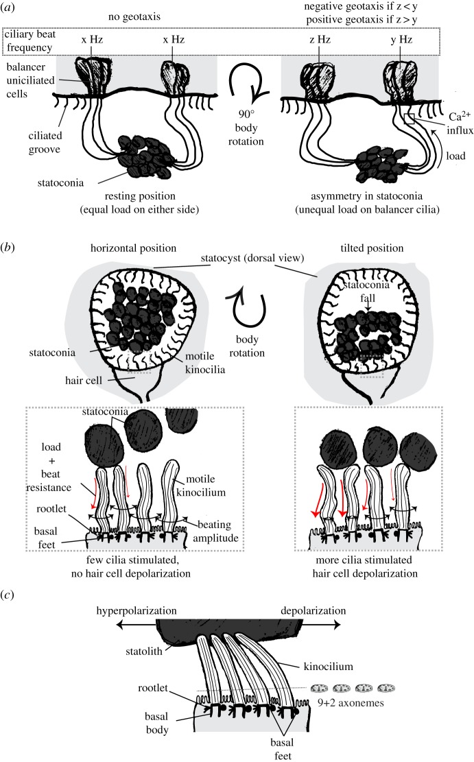
Figure 4.
Diversity in function and structure of ciliated statocysts. (a) (Left) Sketch of a ctenophore statocyst in its resting position (i.e. animal in vertical position). Water surface is to the top. Balancer cilia on opposite sides have the same beating frequency (x Hz). (Right) Upon body rotation the statoconia attached to the balancer cilia exert more load on one group of balancers (arrow). For clarity, the statocyst is not rotated in the sketch. This leads to an asymmetry in stimulation, and thus a consequent difference in beat frequency (z and y). The sign of geotaxis depends on the relative beating frequencies of each balancer. Beating only occurs at the base of the cilia (square) and it depends on Ca2+. (b) (Upper left) Sketch of a non-cephalopod mollusc statocyst in its resting position (i.e. animal in horizontal position). View from the dorsal side. (Lower left) Close-up view of a multiciliated hair cell in resting position. Basal feet are not oriented in the same direction. Some of the motile kinocilia sporadically contact statoconia, thus affecting their beating amplitude. This leads to an increase in voltage noise in the cell, but still below the firing threshold. (Upper right) Upon tilting, statoconia redistribute to one side, thus increasing the chance of cilia colliding with them. (Lower right) More cilia make contact with statoconia, causing depolarization of the cell. (c) Sketch of a cephalopod multiciliated hair cell. Kinocilia are not motile and are attached to the overlying statolith. All basal feet are oriented in the same direction, which is perpendicular to the central pair of the 9+2 axoneme. The cell is depolarized when the cilia are deflected in the direction towards the basal feet and hyperpolarized when bent in the opposite direction. Sketches in a, b and c are based on [184,212], [195,213,214] and [197,215], respectively. (Online version in colour.)

