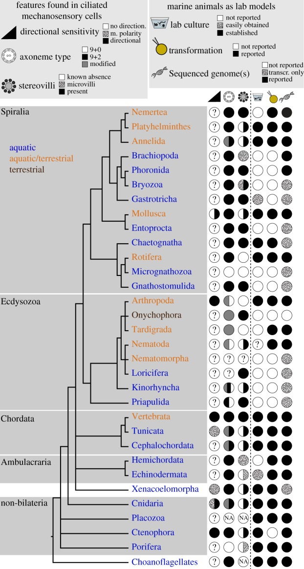
Figure 6.
Animal phylogeny shown to indicate main features of ciliary mechanosensory cells and the degree of current experimental accessibility of each major taxon. First column: evidence of directional sensitivity, morphological polarity (m. polarity) or of no directionality. Second column: type of axoneme found in mechanosensory cells. Third column: presence/absence of cells with stereovilli. Fourth column: marine species that can be reared in the laboratory (dotted circle: a species cannot be breed in the laboratory but can be regularly obtained from the wild). Fifth column: transformation techniques available (e.g. microinjection or electroporation). Sixth column: genome sequence reported (dotted circle: only transcriptome assembly reported). Note that other groups of marine organisms not covered in this review (e.g. Rotifers, Platyhelminthes) also offer good opportunities for experimental analyses. Phylogeny is based on [359–361]. See electronic supplementary material, table S1 for references and further details. (Online version in colour.)

