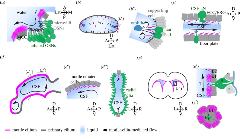
Figure 2.
Schematic depiction of various cavities of the nervous system lined with motile cilia. (a) The olfactory organ of a zebrafish larva is composed of multiciliated cells (MCC) located at the outer rim of the nasal cavity. MCC bear multiple motile cilia (magenta), which generate a directional fluid flow of water. Ciliated OSNs (green), which bear multiple primary cilia (black) and microvilli OSN (grey) are located at the bottom of the nasal cavity. (b,b′) The otic vesicle of a zebrafish embryo at 18–24 hpf contains hair cells (green), or tether cells, that bear primary cilia capable of tethering the otolith (blue). Next to hair cells, there are motile cilia on supporting cells that generate a rotational flow near the otolith. (c) The central canal of the spinal cord is composed of cerebrospinal fluid-contacting neurons (CSF-cNs; green), which bear a microvilli tuft and a motile cilium in zebrafish. ECC (grey), also known as ERG in zebrafish, are located on the floor plate or the dorsal wall of the central canal and bear a cilium. Note that there are more motile cilia in the ventral part of the central canal than the dorsal plane at early developmental stage, and that the CSF flow is bidirectional. (d) The brain ventricular system of the zebrafish larva is decorated by motile cilia (magenta) at very specific locations along the midline. Motile-cilia-mediated flow is complex and compartmentalized to individual ventricles. (d′) Sagittal view of the inset in (d) showing that cells bear a single cilium oriented anteriorly in the same direction as fluid flow. (d″) Transverse view shows that motile cilia are located in the ventral and dorsal wall of the diencephalic-tectal ventricle. Elsewhere, radial glia (RG, green) project their primary cilium into the CSF-filled cavity. (e) ECs, which bear motile cilia, are located along the medial and lateral wall of the mouse lateral ventricle. (e′) Transverse section through the inset in (e) reveals that NSCs of the SVZ are located directly under the ependyma layer made of multiciliated E1 cells and bi-ciliated E2 cells. NSCs also known as B cells (green) project their primary cilium towards the CSF-filled ventricle in addition to contacting the blood vessel (blue), while transient amplifying cells (C cells, grey) and migrating neuroblasts (A cells, grey) lose their direct interaction with the CSF. En face representation shows the pinwheel structure composed of E1 and B cells. Note the translational polarity of the motile cilia of E1 cells. A, anterior; P, posterior; L, left; R, right; D, dorsal; V, ventral; M, medial; Lat, lateral. Motile cilia are in magenta, primary cilia are in black.

