Figure 1.
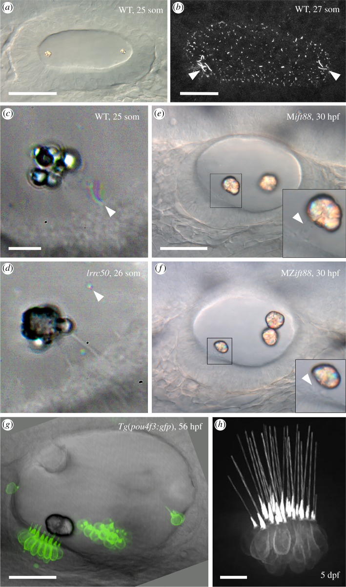
Cilia and otolith formation in the zebrafish ear. (a) Differential interference contrast image of the wild-type otic vesicle at the 25-somite stage, showing the two birefringent otoliths. Scale bar, 40 µm. (b) Lumen of the otic vesicle at the 27-somite stage, stained with an anti-acetylated Tubulin antibody. The arrowheads mark longer tether kinocilia and motile cilia at the anterior and posterior poles of the otic lumen. Scale bar, 20 µm. (c) Time-to-colour merged image of the nascent otolith in a phenotypically wild-type embryo at the 25-somite stage (sibling of the embryo in d). The colour (arrowhead) indicates a motile cilium near the otolith. Scale bar, 10 µm (also applies to d). (d) Time-to-colour merged image of the nascent otolith in a homozygous lrrc50 (dnaaf1) mutant. The otolith has adhered to the tips of two kinocilia (clearly visible in this image), despite the lack of ciliary motility. The arrowhead marks an otolith precursor particle. (e) Phenotypically wild-type embryo (sibling of the embryo in f) lacking maternal ift88 contribution but with normal zygotic ift88 function at 30 hpf. Two otoliths are present, attached to kinocilia (arrowhead, inset). Scale bar, 30 µm (also applies to f). (f) Typical otolith phenotype for a ciliary mutant, in this case an embryo lacking both maternal and zygotic ift88 function. Three otoliths are present, but still localize to the anterior and posterior poles of the ear. The anterior otolith sits directly on the hair cell stereociliary bundles (arrowhead, inset); all cilia are absent. (g) Transgenic zebrafish otic vesicle at 56 hpf expressing GFP in hair cells. Note that the kinocilia of hair cells in the utricular macula do not all contact the anterior otolith (bottom left). (h) Image of a crista in the same transgenic line at 5 dpf. Note the long straight kinocilia. Scale bar, 10 µm. a, b, e, f and g are lateral views with anterior to the left. Abbreviations: dpf, days post fertilization; hpf, hours post fertilization; M, maternal; MZ, maternal-zygotic; som, somite stage; WT, phenotypically wild-type. a, c, d and e are reproduced from [5]; b and f are reproduced from [2]; g is reproduced from [6]; h is reproduced from [7].

