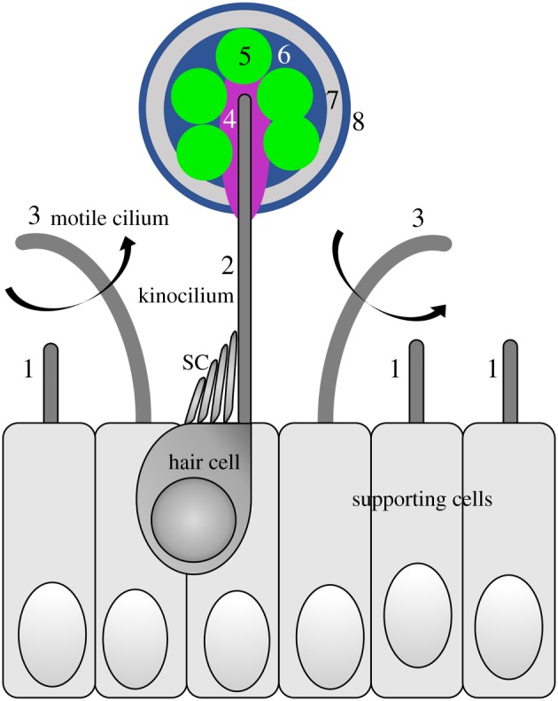
Figure 2.

Schematic diagram summarizing the main early steps in zebrafish otolith formation discussed in this review. A single sensory hair cell is shown for simplicity, but tether cells normally form in pairs (figure 1d). Not to scale. SC, stereocilia. For details and supporting references, see the text. Key: 1, short, immotile cilia, present on most otic epithelial cells; 2, tether kinocilium, present on each of the first sensory hair cells to develop in the ear; 3, motile cilia—these contribute to the accuracy of otolith precursor particle tethering; 4, extracellular proteins, possibly bound to the kinociliary tip, required for otolith precursor particle tethering: Otogelin (and others?); 5, otolith precursor particles, thought to contain Cadherin11, Otoconin90 and Starmaker; 6, otolith proteins incorporated after initial nucleation, including Otolith Matrix Protein-1, Sparc, Starmaker and others; 7, biomineralization: deposition of crystalline calcium carbonate, requiring the activity of Starmaker and Otopetrin 1; 8, further layers added daily throughout life.
