
Figure 2.
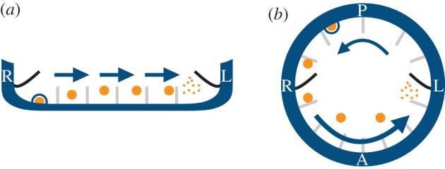
Vesicular chemosensing in (a) the mouse node and (b) the zebrafish KV. A directional cilia-driven flow (arrows) could potentially transport morphogen-filled vesicular parcels (NVPs, orange circles) towards the left where an as-yet unknown active mechanism would break the parcels asymmetrically, releasing morphogen molecules to be sensed by symmetrically distributed cilia. Moreover, the release of NVPs (KVVPs) into the node (KV) might be mediated by shear-induced exocytosis at the walls (blue circle). (L, left of the embryo; R, right; A, anterior; P, posterior). (Online version in colour.)