
Figure 3.
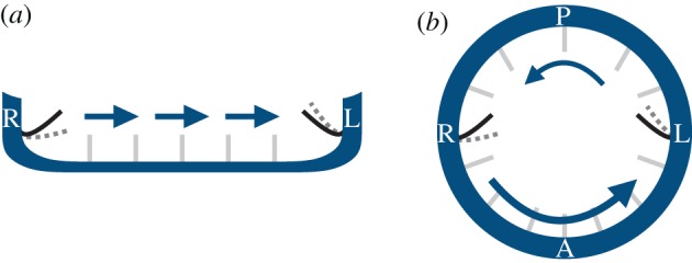
Mechanosensing in (a) the mouse node and (b) the zebrafish KV. Owing to their finite size, cilia from the left/right side of the node (KV) could potentially bend differentially (grey dotted lines) when exposed to a directional cilia-driven flow (arrows). (L, left of the embryo; R, right; A, anterior; P, posterior). (Online version in colour.)