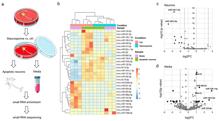
Figure 1.
Analysis of miRNA release from injured cortical neurons into the extracellular space by small RNA sequencing. (a) Flow chart of apoptosis induction. Murine primary cortical neurons from C57BL/6 mice were incubated with either 1 µM staurosporine or DMSO (solvent control) for 8 h. miRNAs were isolated from the resulting supernatants (media) or apoptotic neurons followed by small RNA enrichment and small RNA sequencing (n = 3). (b) Heat map of significantly dysregulated miRNA species. Color scheme depicts Z-Score for each sample. (c) Volcano plot depicting significantly differentially present miRNAs in injured neurons (black dots: fold change FC > ±1.5; p < 0.05). (d) Volcano plot depicting significantly differentially present miRNAs in supernatants of apoptotic neurons (media; black dots: FC ≥ ±1.0; p < 0.05).

