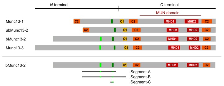
Figure 1.
Domain structure of Munc13 proteins. All Munc13 isoforms share a conserved C-terminal priming unit and a central regulatory unit. Munc13-1 and ubMunc13-2 have a homologous N-terminal region containing an additional C2 domain. CaM-binding sites (dark green bars) are located in the regulatory unit. In bMunc13-2 and Munc13-3, another candidate CaM binding site (light green bars) was initially identified by computational methods, but found to be deficient of CaM-binding in the protein context [28]. C1 = diacylglycerol binding domain (yellow), C2 = Ca2+-binding/protein interaction domain (orange), MHD = Munc homology domain (red). The bMunc13-2 segments investigated in this study are displayed. Segment-A (aa 367–780), segment-B (aa 367–903), and segment-C (aa 703/704–742).

