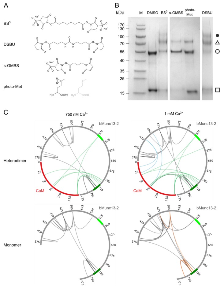
Figure 3.
Investigation of CaM/bMunc13-2 segment-A complex by combining various cross-linking reagents. (A) Structures of applied cross-linking reagents (B) SDS-PAGE (4–20% gradient gel, Coomassie-stained) analysis of complex formation fixed by different cross-linking reagents at 1 mM Ca2+. (M) Prestained Protein Ladder, (□) CaM, (○) segment-A, (∆) CaM/segment-A 1:1 complex, (●) potential CaM/segment-A 2:1 complex. (C) Circos plots showing the identified cross-links with photo-Met (green), s-GMBS (blue), DSBU (dark gray), and BS3 (orange). CaM is illustrated in red and segment-A in gray. For cross-links identified with photo-Met at neighboring Met positions, the most probable one is shown. The established (aa 719–742) and predicted (aa 572–594) CaM-binding sites are shown in dark and light green, respectively (see Figure 1).

