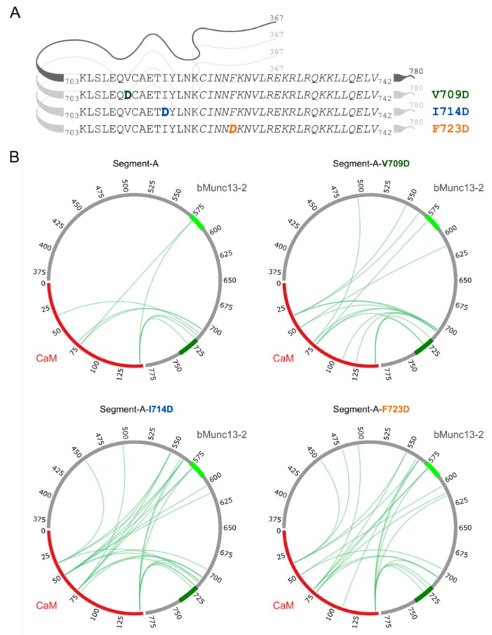
Figure 7.
Segment-A variants and photo-Met cross-linking with CaM. (A) Segment-A variants (V709, I714 and F723) were generated by exchanging hydrophobic amino acids to aspartic acid (bold, colored letters). Displayed are the amino acid sequences of the previously employed segment-C, in which the proposed CaM binding region is shown in italics. (B) Cross-links observed with photo-Met for wild-type and segment-A variants (750 nM Ca2+). CaM is highlighted in red; CaM binding sites are colored in light green (predicted site, aa 572–594) and dark green (established site, aa 719–742) within segment-A (gray).

