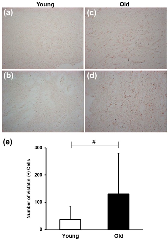
Figure 1.

Age-induced changes in visfatin expression in dental pulp tissues. (a–d) Representative microphotographs of sections of dental pulp stained with anti-visfatin antibodies from individuals aged 17 (a), 20 (b), 35 (c), and 45 (d) years old. The number of visfatin-positive cells increased significantly in the Old group than in the Young group. All microphotographs were taken at an original magnification of × 200. Scale bar: 50 μm. (e) Bar graph illustrating the age-related changes in visfatin immunoexpression in the human dental pulp. Kruskal–Wallis test was performed to analyze the difference in number of visfatin-positive cells between Young group and Old group. # p < 0.0001.
