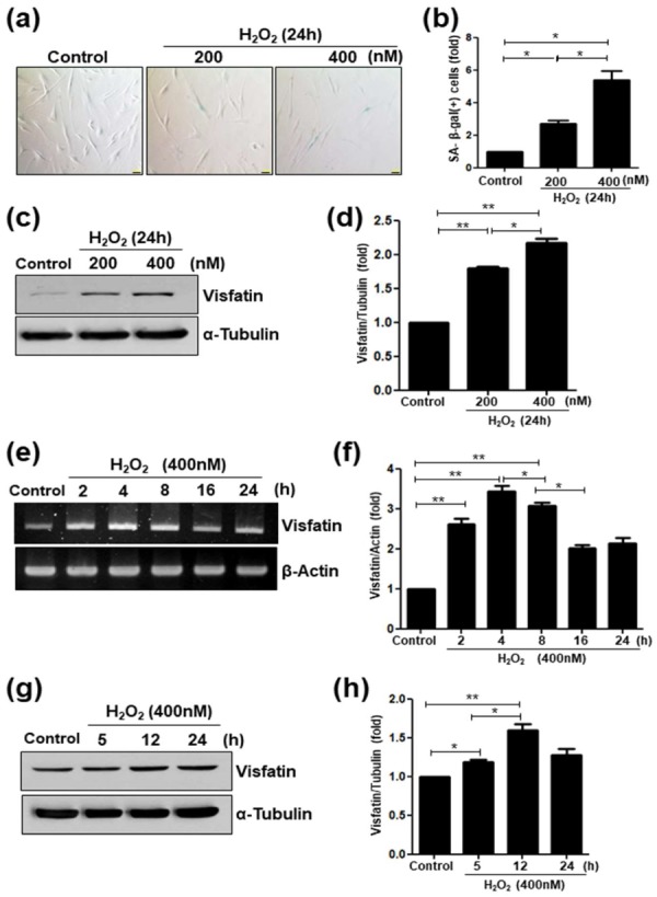
Figure 2.

Upregulation of visfatin in H2O2-induced senescence of human dental pulp cells (hDPCs). (a–d) hDPCs were stimulated with different concentrations of H2O2 (0, 200, and 400 nM) for 24 h. (a) The cells were stained for the detection of the activity of senescence-associated (SA)-β-galactosidase. Scale bar: 200 μm. (b) Quantitative results for the percentage of SA-β-galactosidase positively stained cells. (c) Cells were treated with H2O2 (200 and 400 nM) for 24 h. Cell lysates were subjected to Western blotting for detecting the levels of visfatin or α-Tubulin used as the loading control. (d) Relative visfatin protein levels normalized with α-Tubulin protein levels. (e–h) Cells were incubated with H2O2 (400 nM) for different time periods (0–24 h). (e) Cell lysates were subjected to RT-PCR for determining visfatin mRNA expression. β-Actin was used as an internal control. (f) Relative visfatin mRNA levels normalized with the levels of β-Actin mRNA. (g) Cell lysates were subjected to Western blotting to detect the levels of visfatin protein. α-Tubulin was used as the loading control. (h) Relative visfatin protein levels were normalized with the levels of α-Tubulin protein. * p <0.1, ** p < 0.01.
