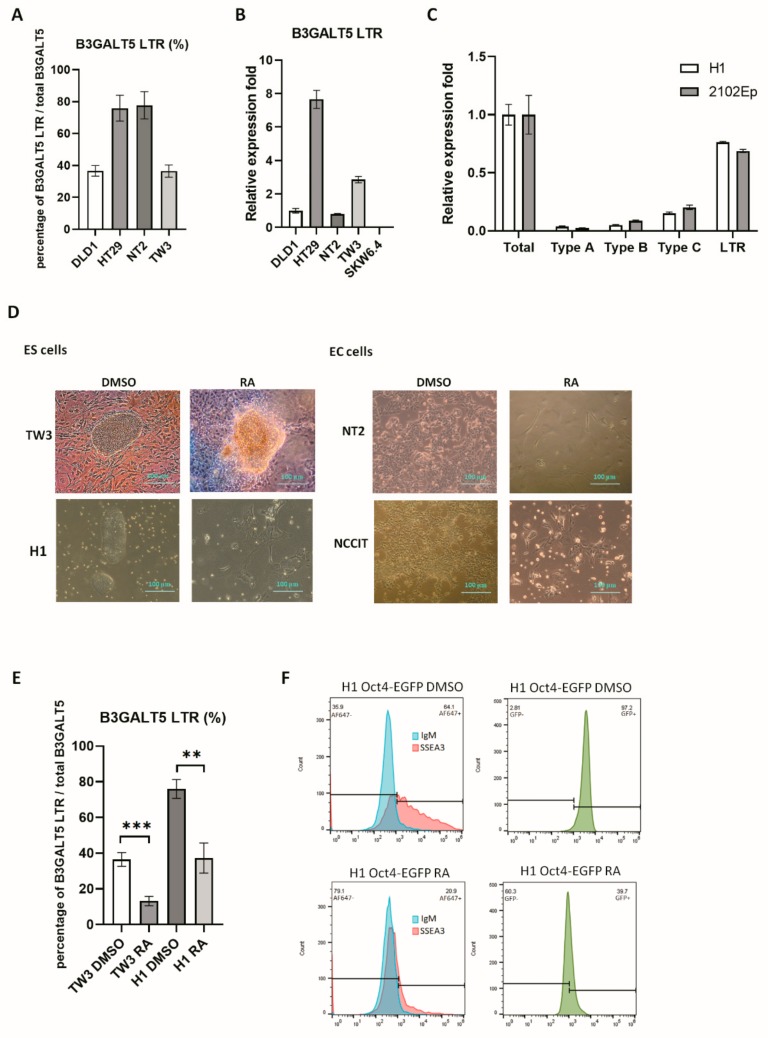
Figure 1.
B3GALT5-long terminal repeat (LTR) expression in embryonic stem (ES) and embryonal carcinoma (EC) cell lines. (A) The expression of B3GALT5-LTR compared with that of total B3GALT5 in colon cancer cell lines (DLD1, HT29), an EC cell line (NT2), and an ES cell line (TW3), reported as a percentage. (B) Normalized expression of B3GALT5-LTR in the cell lines is shown in Figure 1A and in a B-lymphoid cell line (SKW6.4). The DLD1 cell line amount was set to one. (C) Normalized expression of all B3GALT5 transcripts in H1 and 2102Ep cells. Specific primers designed to amplify the B3GALT5 coding region were used to quantify the total transcripts. Three native promoters with their own specific exon were used to design specific primers to quantify as Type A-C transcripts. B3GALT5-LTR transcript levels were higher than Type A-C transcripts. (D) Morphology of the undifferentiated and the retinoic acid (RA)-induced differentiated ES and EC cell lines. (E) Reduction of B3GALT5-LTR expression in ES cells (TW3 and H1) upon differentiation. (p < 0.05; **, p < 0.01; ***) (F) Octamer-binding transcription factor 4 (Oct-4)-controlled expression of enhanced green fluorescent protein (EGFP) was reduced after RA treatment for one week in H1 Oct4-EGFP cells. Stage-specific embryonic antigen-3 (SSEA3) was also decreased after RA treatment.

