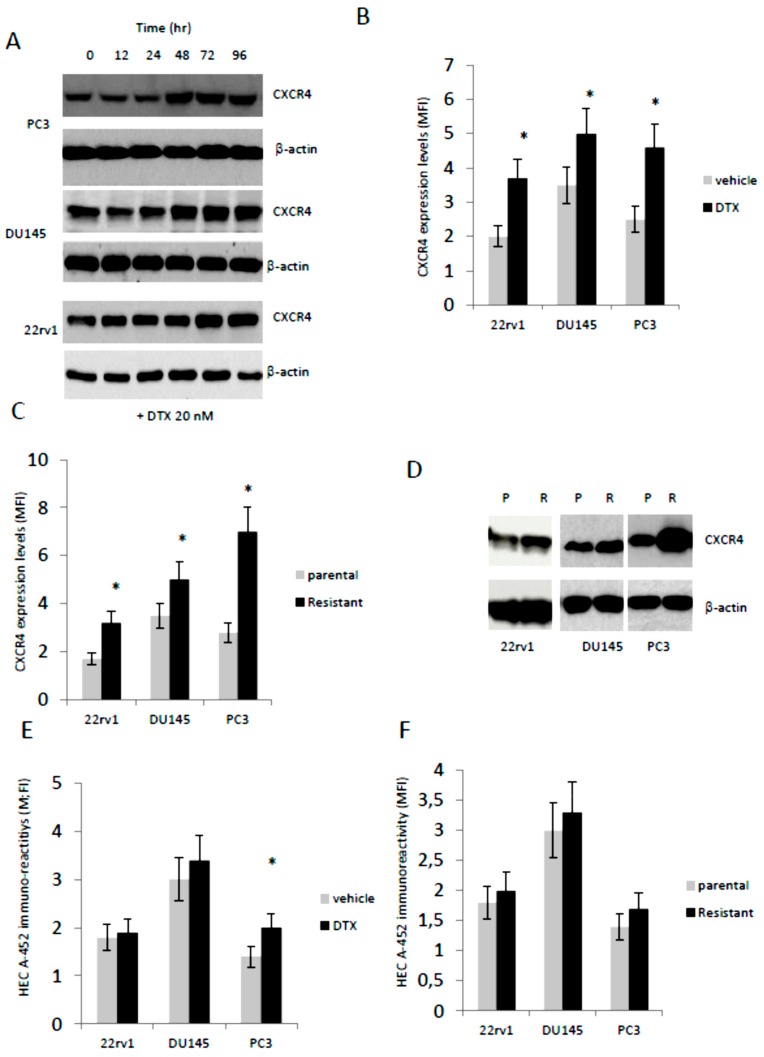
Figure 2.
CXCR4 expression and HECA-452 immune-reactivity after treatment with non-toxic DTX doses. (A) Western blotting analysis performed on PC3, DU145 and 22rv1 cells cultured with 20 nM DTX (time course experiment). (B) Fluorescence analyses on 22rv1, DU145 and PC3 cells cultured with DTX for 96 hr. (C) CXCR4 expression levels by FACS (fluorescence index) on parental and resistant PCa cells. (D) Western blotting analyses performed on parental (P) and DTX resistant (R) 22rv1, DU145 and PC3 cells. (E) HECA-452 immuno-reactivity in parental cell treated with DTX and (F) DTX-resistant cells. Western blots were loaded with 40 µg/lane of proteins. Western blots images are representative o three different gels/experiments. MFI values were calculated for each cell line as indicated in MM ± standard deviation calculated from individual three FACS analyses. * p < 0.05 versus respective controls.

