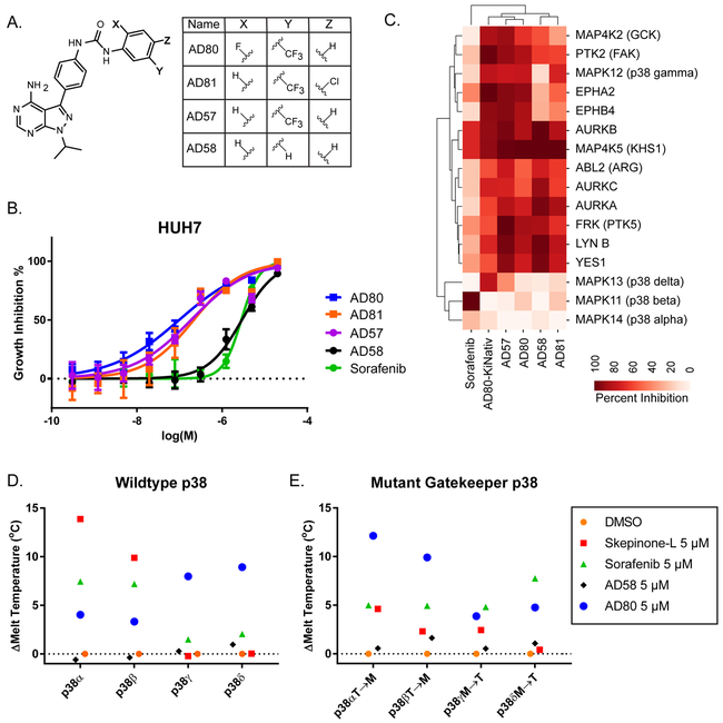
Figure 6: AD80 is isoform biased for p38γ and p38δ, over p38α and p38β, with binding dependent on the gatekeeper residue.
A. Chemical structure of AD80 and related compounds. B. Cell viability assay comparing activity of each compound on the HUH7 cell line. Technical replicates n=6. C. Hierarchical clustering of previously generated in vitro inhibitory profiles (ref. (28)) of the most different kinases between the related compounds. D-E. Melting temperature binding assay using a 1:1 ratio of drug to protein (ie. 5 μM drug to 5 μM protein). Each point is the difference in mean melting temperature (technical replicates: n=6) as determined by differential scanning fluorimetry. In E the following mutations at the gatekeeper residue were tested: p38α/MAPK14 T106M, p38β/MAPK11 T106M, p38γ/MAPK12 M109T, p38δ/MAPK13 M107T.

