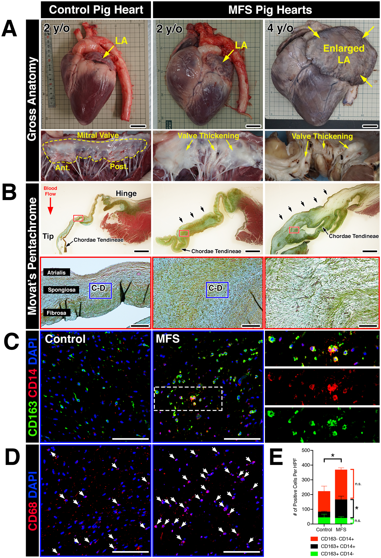
FIGURE 3: Myxomatous mitral valves from Fbn1Glu433AsnfsX98/+ gene-edited pigs with Marfan syndrome exhibit increased monocyte-derived macrophages localized to regions of abnormal ECM remodeling.

A: Whole hearts from wildtype and MFS pigs (Fbn1Glu433AsnfsX98/+) at 2- and 4-years-of-age. Note that the left atrium is enlarged in the MFS heart at 4 years-of-age (yellow arrows). Corresponding mitral valves including anterior and posterior leaflets are depicted in situ. Valve thickening is apparent by gross inspection, illustrated by increased opaqueness of the leaflets versus wildtype controls (yellow arrows). B: Histological sections of posterior leaflets of the mitral valves stained with Movat’s pentachrome are depicted and show thickening (arrows; upper panels) of the mitral valves in MFS pig hearts at 2- and 4- years-of-age versus controls. Note that the chordae tendineae are also markedly thickened in MFS hearts. At higher magnification (lower panels), MFS pig hearts exhibit loss of tri-laminar ECM organization of the atrialis (elastin), spongiosa (proteoglycan), and fibrosa (collagen) layers versus controls. C: By immunofluorescence, CD14+ monocytes and CD163 macrophages can be observed in the regions highlighted by the dotted box (B, lower panels). Overall, monocytes and macrophages were increased in Marfan syndrome pigs vs wildtype controls. Magnified view of double-positive cells is depicted in insets and separated by red and green fluorescent channels. D: CD68+ macrophages are depicted in comparable regions using sister sections. E: Quantification of macrophages in MFS pigs (n=6) versus controls (n=3). Data are represented as mean ± SEM. *p<0.05. n.s. = not significant. Scale bars = 300mm (A); 2mm (upper panels in B) and 200μm (lower panels in B); and 100μm (C, D). Abbreviations: LA=Left atrium; y/o=year-old; MFS=Marfan Syndrome; Ant.=Anterior leaflet; Post.=Posterior leaflet; HPF=High power field.
