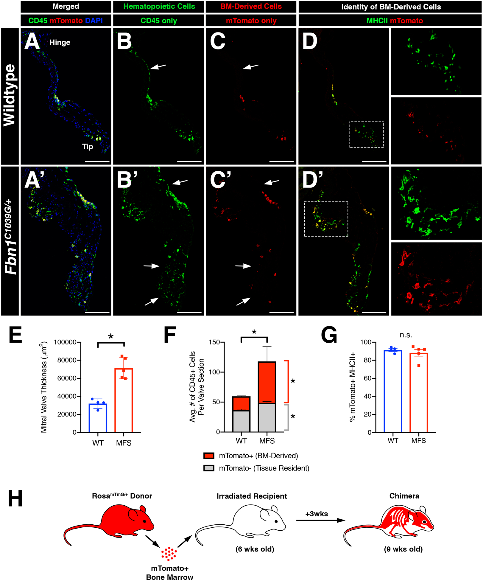
FIGURE 5: Inflammatory micro-environment in myxomatous valves recruits bone-marrow-derived monocytes that contribute to one of two distinct macrophage populations in the heart valve.

Infiltrating CD45+ cells populate the valve leaflets during homeostasis and disease. A-H: Bone marrow (BM) transplantation was performed on irradiated 6 week-old Fbn1C1039G/+ mice and age-matched controls and analyzed 3 weeks later. Immunofluorescence demonstrates the presence of mTomato+ donor cells in the mitral valve (red; C, C’) which are also CD45+ (green; B, B’; yellow; A, A’). Note the presence of host-derived CD45+ cells in merged panels (green; A’, A’). mTomato+ donor cells were predominantly MHCII+ (D, D’). E: Mitral valve leaflets remained thickened in Marfan syndrome mice versus controls after BM transplant, as expected. F: Marfan syndrome mice exhibit increased BM-derived CD45+ cells versus controls. Tissue-resident CD45+ cells also show a modest increase in number in Marfan syndrome mice. G: Quantification of MHCII+ cells among mTomato+ cells in the mitral valve indicates that BM-derived cells are predominantly MHCII+. H: Schematic of experiment. Data are represented as mean±SEM. *p<0.05. n.s. = not significant. n=4–5 mice/genotype. Scale Bar = 100μm. Abbreviations: BM=Bone marrow; MFS=Marfan Syndrome; wks=weeks.
