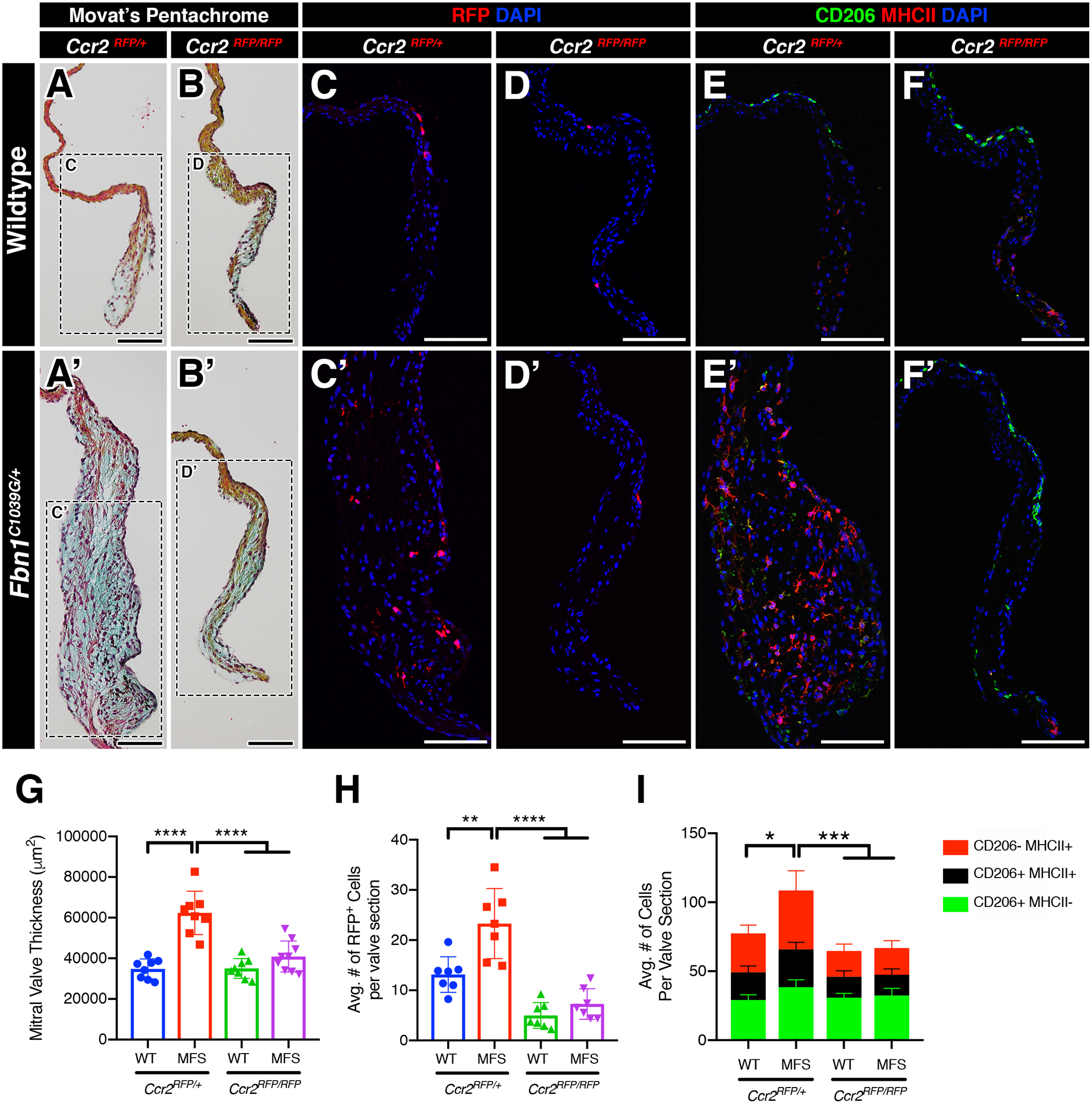
FIGURE 6: Deficiency of infiltrating CCR2+ monocytes inhibits the progression of myxomatous degeneration in Marfan syndrome mice.

A-B’: Representative Movat’s pentachrome stained sections of the mitral valves (anterior leaflet) in 2-month-old WT and MFS mice in the normal (Ccr2RFP/+; A, A’) and CCR2-depleted (Ccr2RFP/RFP; B, B’) state. Note that the leaflet thickness is notably reduced in MFS mitral valves in the depleted (B’) vs normal (A’) state. C-D’: By immunofluorescence, CCR2+ monocytes (red) are significantly increased in MFS mitral valves (C’) versus WT (C). CCR2-depleted mice exhibit significant reduction in monocytes in both WT (D) and MFS (D’) mitral valves. E-F’: CCR2+ monocytes contribute mainly to the MHC-II macrophage subpopulation and an increase in monocyte population is correlated to the increase in the MHC-II macrophage subpopulation versus controls. Deficiency of CCR2 results in a significant reduction of the MHC-II+ population in both wildtype and MFS mice. G-I: Quantification of mitral valve thickness (G), number of CCR2+ monocytes (H), and number of macrophage subpopulations (I) in WT and MFS mice in the normal and CCR2-depleted state. Data are represented as mean ± SEM. *p<0.05, **p<0.01, ***p<0.001, ****p<0.0001. n=8–9 mice/genotype. Data are represented as mean ± SEM. Scale Bar = 100μm. Abbreviations: WT=Wild Type; MFS = Marfan Syndrome; RFP=Red fluorescent protein.
