Figure 2.

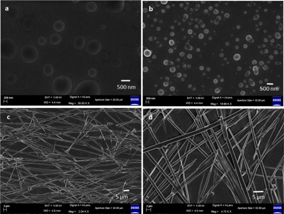
FE-SEM images of (a) peptide 1 showing polydisperse microsphere morphology, (b) peptide 2 showing polydisperse microsphere morphology, and (c,d) peptide 3 showing entangled fiber-like morphology.

FE-SEM images of (a) peptide 1 showing polydisperse microsphere morphology, (b) peptide 2 showing polydisperse microsphere morphology, and (c,d) peptide 3 showing entangled fiber-like morphology.