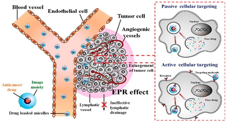
Figure 6.
Schematic representation of drug loaded micelles (spheres) with imaging agents, from the administration site to the tumour tissue. After administration, micelles (10–200 nm) display specific targeting of tumour growth via passive targeting with cellular endocytotic uptake from exterior fluid to the cancer cells. Active targeting through receptor-mediated internalization is achieved by attachment of antibody ligand molecules, to the surface of micelles (Adapted with permission from Chen et al. [81]).

