Figure 4.
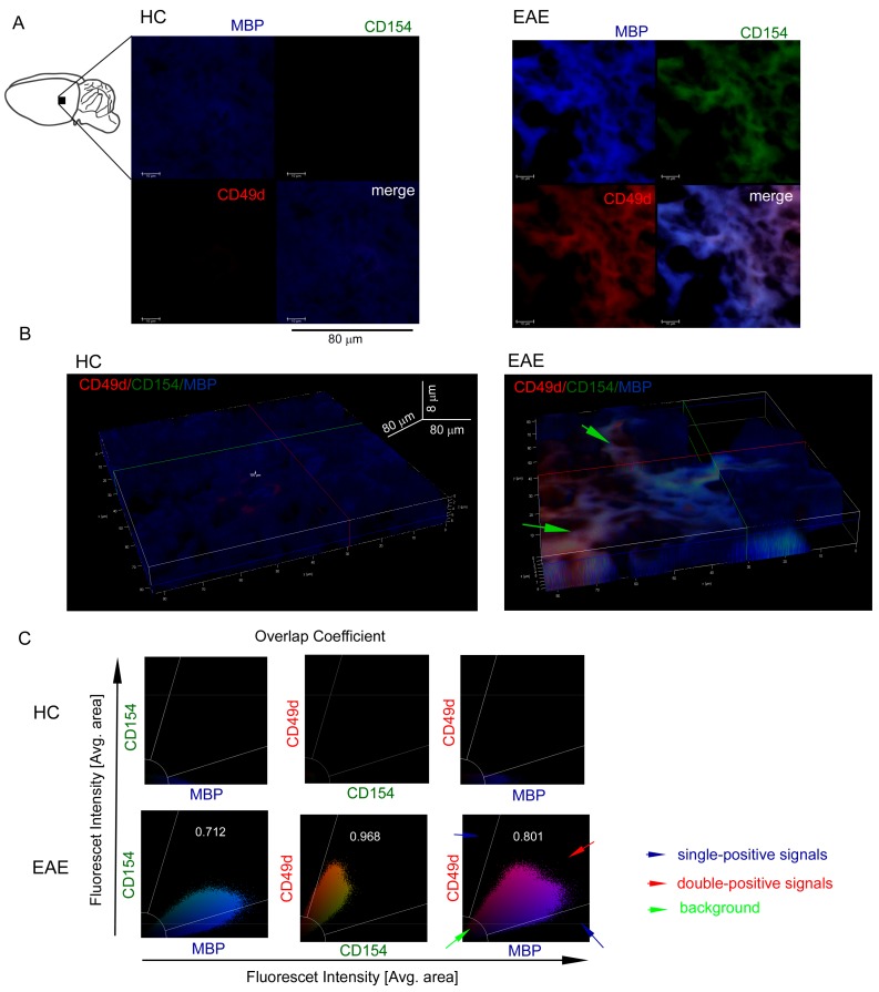
CD49d+CD154+ cells in the EAE brain three weeks after the disease peak are localized close to the remyelination lesions. (A) Immunohistochemistry (IHC) microscopy analysis of sagittal brain slices revealed high MBP+ (blue pseudocolor) signal three weeks after the EAE peak (upper-left picture) confirming remyelination. High MBP+ expression was accompanied by the high CD49d+ (red pseudocolor) and CD154+ expressions (green pseudocolor). The example of one of four independent experiments (EAE n = 4, HC n = 4). (B) Z-stack analysis in 3D projection demonstrated that CD49d+/CD154+ signals were co-localized around the regions with high MBP+ signal. Green arrows point the regions with high double-positive signal for CD49d+ and CD154+ close to the regions with high MBP expression. The example of one of four independent experiments (EAE n = 4, HC n = 4). (C) The example of overlap coefficient analysis (EAE n = 4, HC n = 4). Cytofluorograms of CD154 vs. MBP, CD49d vs. CD154, and CD49d vs. MBP signals from the EAE brain confirmed high expression of MBP with co-expression of CD49d+CD154+ cells.

