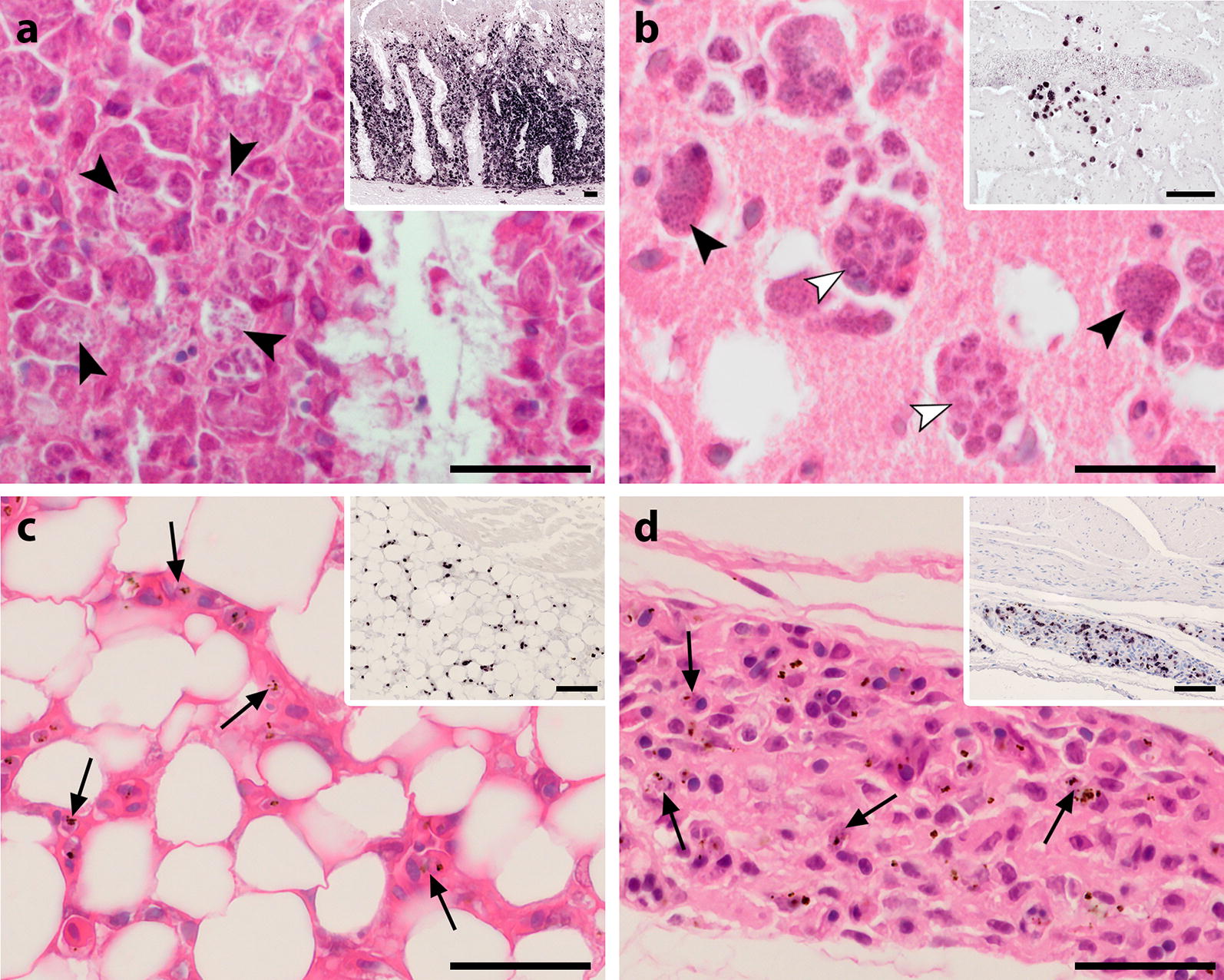
Fig. 2.

Histological sections of the intestine (a), brain (b), heart (c) and gizzard (d) from infected Eurasian blackbirds (Turdus merula) showing developmental stages of Plasmodium matutinum (a, b, d) and P. vaughani (d). Parasites were visualized by CISH (a–d, inserts, low magnification) and identified in haematoxylin–eosin stained tissue sections (a–d, high magnification). a In the intestine, CISH revealed areas of excessive merogony of P. matutinum in the intestinal mucosa (insert). Numerous meronts of P. matutinum (arrowheads) were located in the cytoplasm of large cells (presumably macrophages) of the lamina propria mucosae accompanied by severe displacement and atrophy of crypt structures. b In the brain, clusters of meronts were occasionally observed in several birds, here showing meronts of P. matutinum located close to a large vessel (insert). Some of the meronts showed multiple merozoites-containing compartments (white arrowheads). c, d In a number of birds, abundant small CISH signals were detected in fat tissue (c, insert) and the subserosal layers of visceral organs (d, insert). In the corresponding HE-stained sections, erythrocytic parasite stages (arrows) were observed to accumulate in these tissues. Scale bars: 25 µm, inserts: 50 µm
