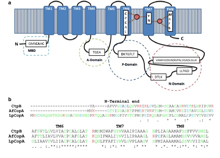
Fig. 1.
Topology, functional motifs, and domains of M. tuberculosis CtpB. a The predicted topology of CtpB shows eight TM helices (1 to 8), locating the N- and C-terminal ends within the cytoplasmic portion. The cytoplasmic domains are represented by stitched lines and the functional motifs of P1B-type ATPase are shown. The amino acids responsible for cation coordination within TM segments 6, 7 and 8 are highlighted in bold. b The TM helices were predicted using TOPCONS. The multiple alignment of the N-terminal end, TM6, TM7, and TM8 of CtpB and other bacterial characterized Cu+-ATPases (CopA from Archaeoglobus fulgidus (WP_010877980.1) and Legionella pneumophila (YP_095057.1)) was constructed using Clustal Omega. Some of the amino acids involved in the coordination of Cu+ are located within TM6, 7 and 8. The residues are classified as nonpolar (black), polar (green), acid (blue), and basic (red)

