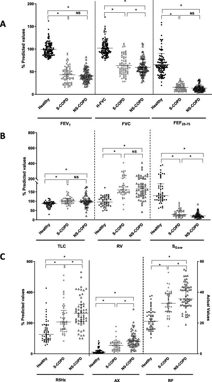
Fig. 1.
Differences in lung function between healthy (n = 114, ●), smoking (S)-COPD (n = 71, ■),) and non-smoking COPD (n = 82, △) using (a) spirometry, with forced expiratory volume in 1 s (FEV1), forced vital capacity (FVC) and forced expiratory flow between 75 and 25% of vital capacity (FEF25–75); b Lung volumes, showing total lung capacity (TLC), residual volume (RV), specific airway conductance (sGaw) and specific airway resistance (sRaw); c Impulse oscillometry, showing resistance at 5 Hz (R5), area under the reactance curve (AX), resonant frequency (RF). Data are presented as individual data points and the bars indicate median and interquartile ranges, where * indicates p < 0.05, NS = non-significant

