Fig. 1.
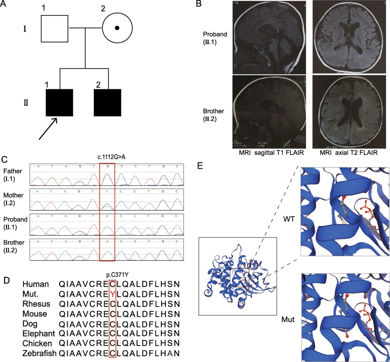
The novel likely pathogenic variant of the PAK3 gene and the brain MRI scans of the patients. a The family tree. b The left sides show sagittal T1 FLAIR images with widened lateral ventricles and thin corpus callosum. The right sides show axial T2 FLAIR images with enlarged lateral ventricles. c The hemizygous variant detected by whole-exome sequencing and confirmed by Sanger sequencing. d The variant region is conserved among human, rhesus, mouse, dog, elephant, chicken and zebrafish. e The predicted structure of the mutated PAK3, with the site of the mutation in the enlarged views

