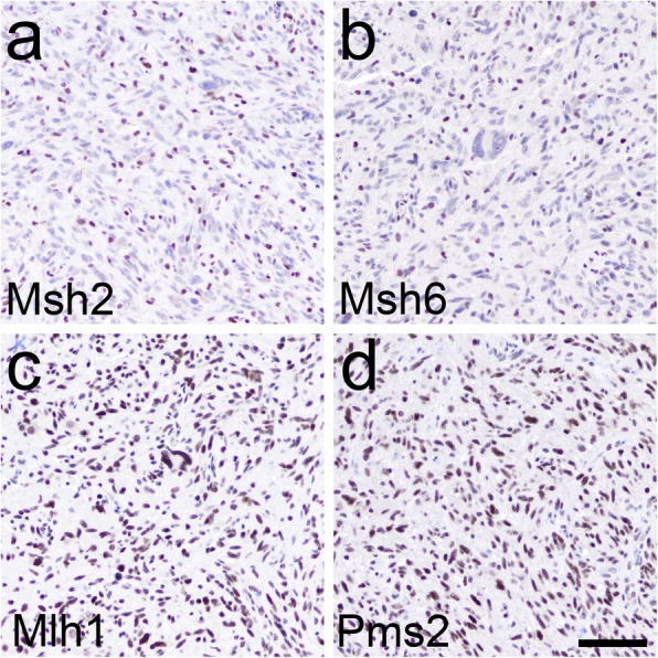
Fig. 1.

MMR IHC in Tumor 1. The tumor was a recurrent GBM, post-TMZ therapy, in a 57 year-old woman (Table 2). Tumor cells showed loss of Msh2 (a) and Msh6 (b), and retention of Mlh1 (c) and Pms2 (d). Note the normal immunostaining within nonneoplastic cells scattered throughout the tumor in (a) and (b). Scale bar = 100 μm
