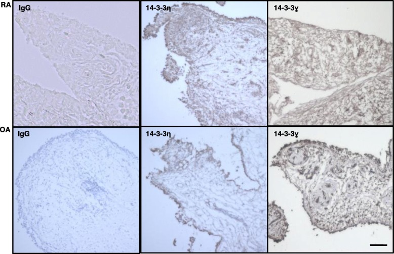
Fig. 1.
Higher expression of 14-3-3η+ and fewer 14-3-3ɣ+ cells were detectable in the RA synovium than in OA synovium. Synovium specimens of patients with RA (n = 3) or OA (n = 3) were incubated with the indicated primary antibodies and then visualized using DAB chromogen. Representative images from three independent experiments are shown. Panels labelled IgG, isotype control. Scale bar, 50 μm

