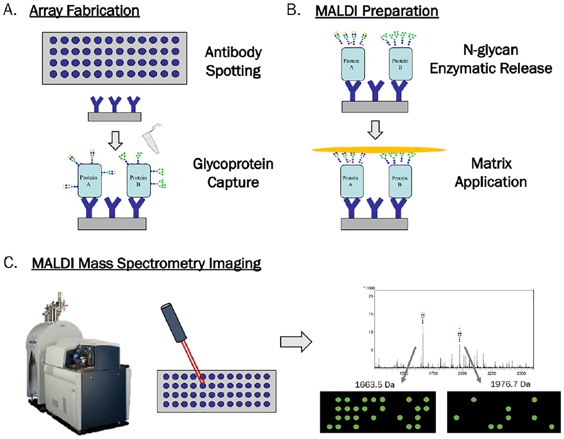
Figure 1.

Workflow for Antibody Panel Based N-glycan imaging by MALDI MS. A) Antibodies are spotted and slide blocked with BSA. Sample is added for capture of glycoproteins by their antibodies. B) N-glycans are enzymatically released in a localized manner followed by matrix application. C) Slides are imaged by a MALDI FT-ICR MS to obtain overall spectrum and individual images for each m/z peak, which show the abundance of each N-glycan in two-dimensions across the array.
