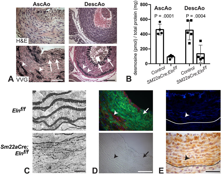
Figure 4. Analysis of the aorta in mice with homozygous deletion of Eln in SMCs.
(A) H&E and VVG staining of P18 ascending (left) and P12 descending (right) aorta show the elastic laminae structure. Sm22aCre;Elnf/f ascending aorta has a fragmented IEL, while the descending aorta has an intact IEL (arrows). Both arteries have patches of elastin (arrowheads) in the outer half of the media near the adventitia. Littermate control images are shown in Figs. 2A and B. Scale bars = 50 μm. (B) Desmosine levels were expressed as a ratio of total protein in the ascending and descending aorta. Control is Elnf/f and Elnf/+ combined. P values were determined between genotypes for each artery by two-way ANOVA with Sidak’s multiple comparisons test. (C) Electron microscopy images of the outer media show that the patches of elastin present in Sm22aCre;Elnf/f ascending aorta are thin and fragmented. Scale bar = 10 μm. (D) Lineage tracing with the ROSA26mT/mG reporter mouse demonstrates Sm22aCre-mediated recombination (green) in the media corresponding with the absence of elastin, as well as in the neointima (arrow). There are also cells in outer regions of the wall near the patches of elastin that do not show Sm22aCre-mediated recombination (red, arrowhead). Bright field image of the same field is shown below for comparison. Scale bar = 50 μm. (E) Patches of elastin are shown with autofluorescence in P2 Sm22aCre;Elnf/f ascending aorta (arrowhead, top). The medial cells stain positive for SM22a protein even if they are near the patches of elastin associated with nonrecombination (arrowhead, bottom). The border between the media and adventitia is indicated. Scale bar = 50 μm.

