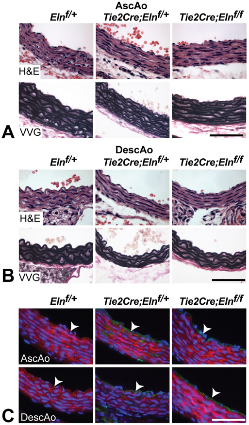
Figure 5. The IEL appears normal in the aorta despite EC-specific Eln deletion.
H&E and VVG staining of P10 ascending (A) and descending aorta (B) show normal IEL and medial lamellae formation despite EC-specific Eln deletion. (C) Lineage tracing with the ROSA26mT/mG reporter mouse confirms green (recombined) ECs in Elnf/+ and Elnf/f mice expressing Tie2Cre and red (not recombined) ECs in Elnf/+ controls (arrowheads). All scale bars = 50 μm.

