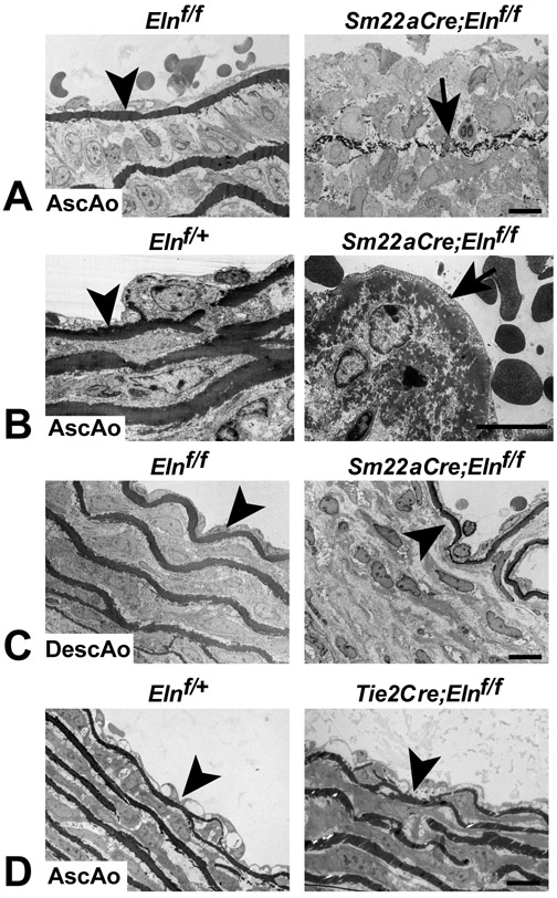
Figure 6. Electron microscopy of the IEL in SMC- and EC-specific Eln knockouts.
(A) The IEL in the ascending aorta of P8 Elnf/f mice is intact (arrowhead), while in some regions it is composed of small aggregates of elastin that never fused to form a continuous barrier in Sm22aCre;Elnf/f mice (arrow). (B) In regions where the IEL is thicker and more intact in Sm22aCre;Elnf/f ascending aorta, it has a moth eaten appearance (arrow). (C) In contrast, the descending aorta in P8 Elnf/f and Sm22aCre;Elnf/f mice has an intact IEL (arrowheads). (D) The IEL is also intact in the ascending aorta of P10 Elnf/+ and Tie2Cre;Elnf/f mice (arrowheads). All scale bars = 10 μm.

