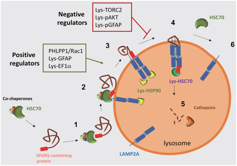
Fig. 1.
Chaperone-mediated autophagy: schematic model of the steps in chaperone-mediated autophagy.1. Substrate binding by HSC70 and cochaperones and targeting to lysosomes. 2. Binding of the substrate to LAMP2A at the lysosomal membrane. 3. HSP90 binds to LAMP2A to stabilize it while it organizes into higher molecular weight complexes. 4. Substrate crosses the lysosomal membrane through a LAMP2A-enriched translocation complex, and translocation is complete by the action of luminal HSC70. 5. The substrate is rapidly degraded by luminal proteases (cathepsins). 6. Once substrate translocation is complete, LAMP2A dissociates into monomers in a process dependent on cytosolic HSC70. Red box: negative regulators of CMA at the lysosomal membrane. Green box: positive regulators of CMA at the lysosomal membrane

