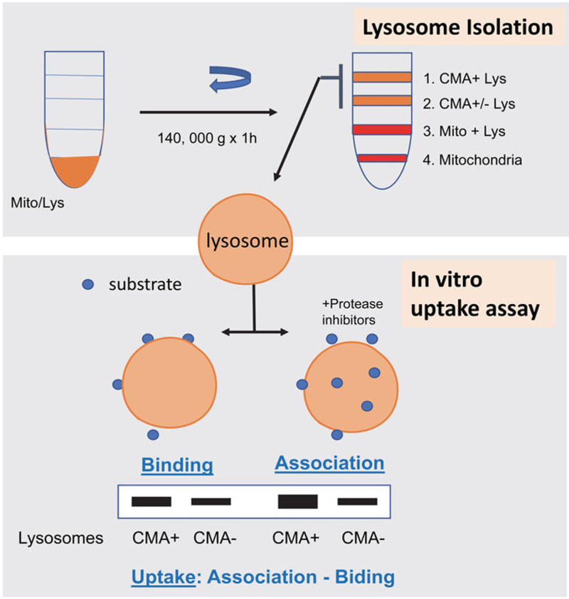
Fig. 2.
Measurement of CMA in vitro. Top: lysosomes active for CMA can be isolated by flotation in a discontinuous gradient of metrizamide. Bottom: incubation of CMA substrates with intact lysosomes pre-treated or not with inhibitors of lysosomal proteases allows to quantify substrate binding and translocation (uptake) inside the lysosomal lumen

