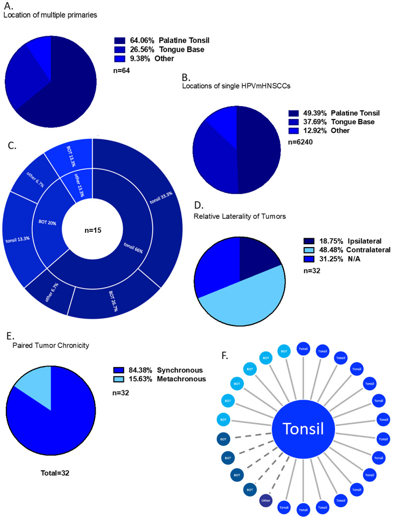
Figure 3. Multiple HPVmHNSCC with at least one primary in the oropharynx from SEER database.
A) Locations of multiple HPVmHNSCC. “Other” refers to tumors located in the hypopharynx, nasopharynx, or oral cavity and not otherwise included in the first two categories. B) Locations of single HPVmHNSCCs. C) Locations of multiple HPVmHNSCC that were diagnosed at different timepoints. The inner ring corresponds with the first diagnosed primary, and the outer ring corresponds with the second diagnosed primary. D) Tumor laterality. E) Frequency of synchronous tumors (defined as diagnosis within 6 months) compared to metachronous tumors (defined as diagnosis more than 6 months apart). F) Sites of second tumors after a first primary located in the tonsil from SEER. Solid line represents tumors identified synchronously and dotted line represents tumors identified metachronously.

