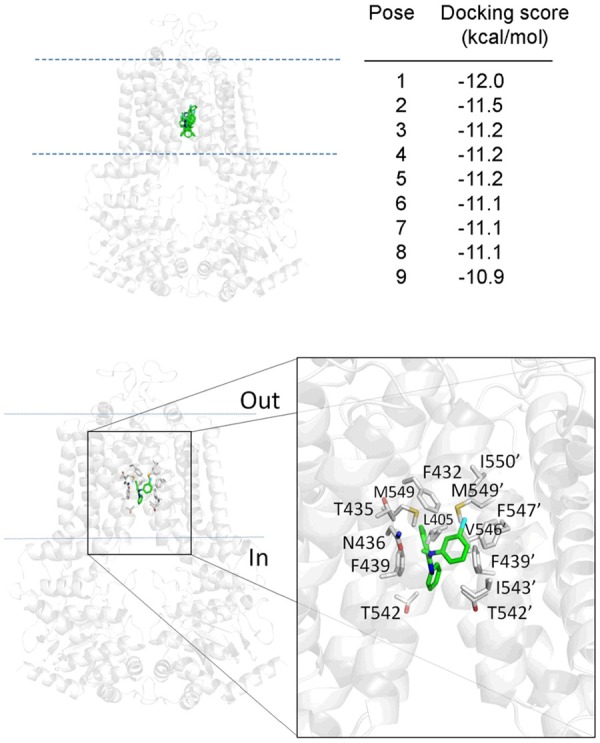
Figure 6.

Binding of MY-5445 in the drug-binding pockets of ABCG2. MY-5445 was docked to the cryo-electron microscopy structure of human ABCG2 (PDB: 5NJ3) using Autodock Vina software as described in Methods. All nine low-energy poses of MY-5445 interaction with residues in the same binding cavity in the transmembrane region (green sticks, top left panel), with similar binding affinities (top-right). The amino acids within 5Å of MY-5445 in the lowest-energy pose are shown with gray sticks (bottom panel). Pymol software was used for analyses of docking poses and figure preparation.
