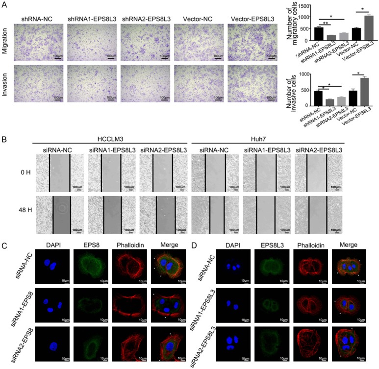
Figure 3.

EPS8L3 promoted cell migration and invasion of HCC in vitro. (A) The migratory and invasive abilities of Huh7 cells were evaluated by transwell-assays. Representative images were shown in left panel. The numbers of migratory or invasive cells in different groups were shown in the right panel. (B) Representative images of wound healing assay. The cell-free gap was much narrower in the negative control group than in the EPS8L3 knockdown groups. (C, D) Representative images showing the changes of F-actin and pseudopodia after EPS8 (C) or EPS8L3 (D) knockdown in Huh7 cells. Results of three independent experiments were shown as mean ± s.d. *: P<0.05, **: P<0.01.
