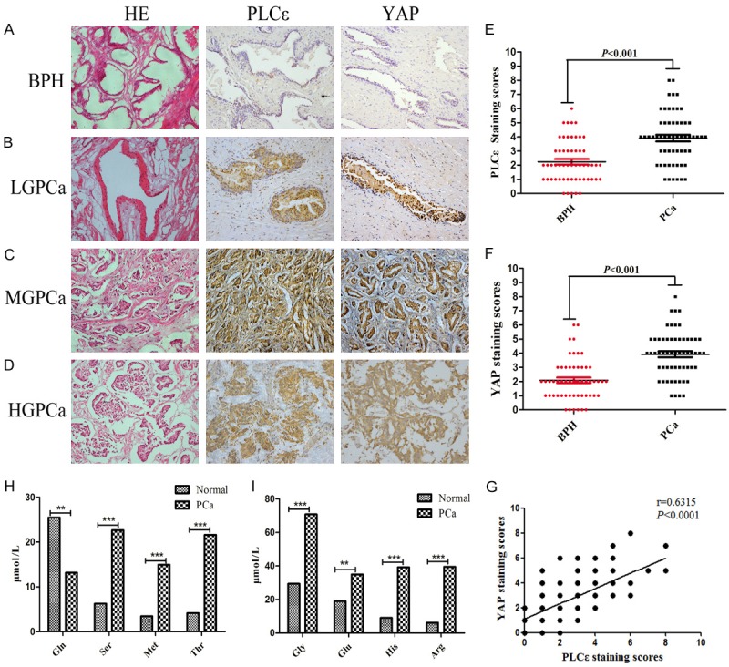
Figure 1.

High expression of PLCε and YAP in tissues and high levels of serine/glycine in blood of PCa. (A-D) Representative hematoxylin and eosin (H&E) staining and IHC staining in 55 PCa and 58 BPH samples. Magnification × 200. Representative IHC staining of different staining intensities was used as a criterion for staining scores: BPH (A): with no staining; Low-grade (LG) PCa (B): with light staining; Middle-grade (MG) PCa (C): with moderate staining; High-grade (HG) PCa (D): with strong staining. (E, F) Staining scores of PLCε (E) and YAP (F) expression in BPH and PCa tissues. Data were represented as the means ± SD. (G) Correlation analysis between PLCε and YAP in tissues analyzed by Pearson analysis. (H, I) Multiple common amino acids concentration levels in different blood samples tesetd by mass spectrometry and analyzed by Mann-Whitney test. *P<0.05, **P<0.01, and ***P<0.001 vs. controls.
