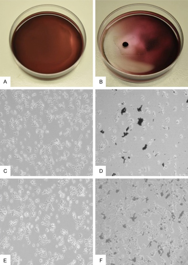
Figure 4.

Magnetic-targeting effect of BLM-MNPs on tumor cell in vitro. A. Representative macroscopic image of Cal-27 cells treated with BLM-MNPs after 0 h of incubation; B. Representative macroscopic image of Cal-27 cells treated with BLM-MNPs after 0 h of incubation under magnet field. BLM-MNPs clustered around the magnet, manifesting as reduced cytotoxicity to normal cells at this area with the drug loaded on it; C. Representative microscopic image of Cal-27 cells treated with BLM-MNPs after 24 h; D. Representative microscopic image of Cal-27 cells treated with BLM-MNPs after 24 h of incubation under magnet field, the number of cells declined considerably in magnetic field (Black: BLM-MNPs); E. Representative microscopic image of Cal-27 cells treated with BLM-MNPs after 48 h of incubation; F. Representative microscopic image of Cal-27 cells treated with BLM-MNPs after 48 h of incubation under magnet field (Black: BLM-MNPs).
