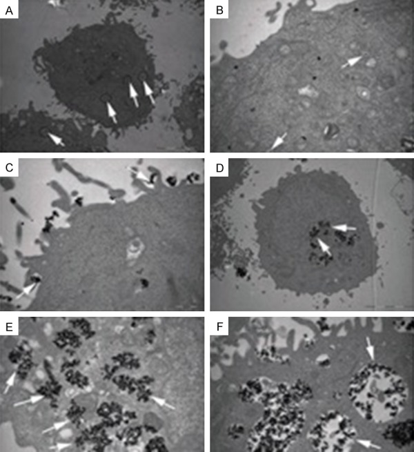
Figure 5.

TEM images of Cal-27 treated with BLM-MNPs after 24 h of incubation. A. The BLM-MNPs were observed mainly in the cytoplasm (white arrows indicate diffusive nanoparticles, scale bar: 5 µm); B. The BLM-MNPs were found to be located inside the endosome (white arrow, scale bar: 5 µm); C. Cal-27 cells formed pseudopodia to endocytose BLM-MNPs (white arrow, scale bar: 1 µm); D. A great number of BLM-MNPs were intruding into the nucleus of Cal-27 cells (white arrow, scale bar: 1 µm); E, F. A great number of BLM-MNPs were situated inside the vesicles (white arrow, scale bar: 1 µm).
