Figure 7.
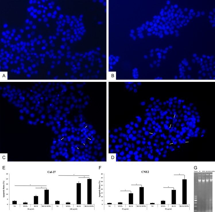
Apoptosis assay of BLM-MNPs on tumor cell in vitro. (A-D) Localization of nucleus, which was stained with 4’,6-diamidino-2-phenylindole (DAPI). In the NS group (A), the nucleus was homogeneously stained and no DNA damage was observed. The nuclear blue fluorescence was also clear in the MNPs group (B). After treatment with BLM (C) and BLM-MNPs (D), the characteristics of apoptosis, such as nuclear fragmentation and chromatin condensation, were observed in the nucleus (C and D, white arrow); (E, F) The effect of BLM-MNPs on apoptotic rate of Cal-27 and CNE2 cells were detected by flow cytometry in comparison with NS, MNPs and BLM. 24 h of treatment with MNPs resulted in few apoptotic cells. The apoptotic rates in BLM-MNPs treated cells were significantly higher in contrast to BLM treated cells, and in BLM-MNPs treated cells were significantly higher in contrast to NS or MNPs treated cells (*P<0.05). These results have clearly demonstrated that BLM-MNPs has the apoptosis effect on Cal-27 and CNE2 cells. (G) After incubation for 24 h, characteristics of apoptosis (DNA ladder bands) were detectable in BLM-MNPs and BLM groups. No DNA fragmentation was detected when cells were exposed to MNPs or NS.

