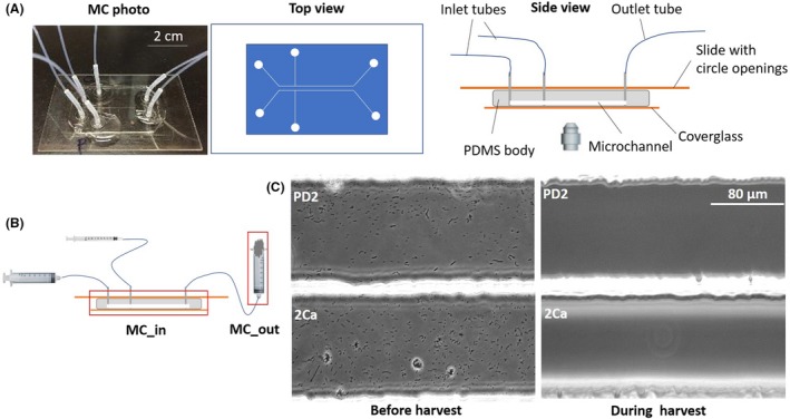
Figure 1.

Preparation of X. fastidiosa cultures in microfluidic chamber (MC) for whole transcriptome analysis.
A. MC with dual parallel channel design. Left picture shows an assembled MC, middle diagram shows top view, and right diagram shows side view of the MC.
B. Side view of the MC during X. fastidiosa growth experiments. 5 ml glass syringes filled with media were connected to medium inlets, 1 ml plastic syringes filled with X. fastidiosa suspension were connected to bacterium inlets, and 10 ml plastic syringes sealed with cotton balls were connected to outlets. During cell harvest, 1 ml plastic syringe filled with RNA/DNA Shield buffer replaced the 5 ml glass syringes, and the 10 ml plastic syringes were replaced by 1.5 ml microcentrifuge tubes.
C. Micrographs of the two parallel channels showing the growth of X. fastidiosa and harvest of cells from channels.
