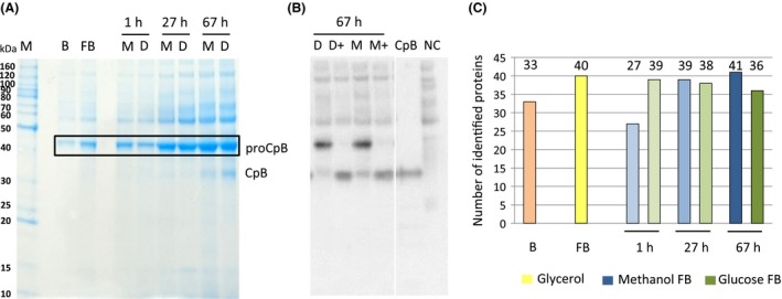
Figure 3.

Analysis of proteins present in the culture supernatant.
A. Exemplary SDS‐PAGE of culture supernatants of the glycerol batch phase (B), glycerol fed‐batch (FB) and different time points of the glucose (D) and methanol (M) fed‐batch processes (1, 27 and 67 h; A).
B. Anti‐CpB Western Blot of cultures (1:10 diluted culture supernatants, reduced and denatured), probed with monoclonal HRP‐conjugated anti‐CpB antibody (1:1500) and detected by chemiluminescence; + indicates that samples were treated with trypsin to activate pro‐CpB to mature CpB.
C. Number of identified proteins in culture supernatants of the corresponding samples shown in A.
