. 2019 Aug 20;10(39):8930–8938. doi: 10.1039/c9sc02906k

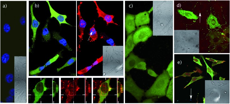
Fig. 3. Transport experiments in cells. (a) Control experiments in HeLa cells showing confocal micrographs of cells incubated with pyranine (5 μM) and cage C (5 μM); nuclei stained with Hoechst (blue). (b) Top: confocal micrographs of HeLa cells incubated with both pyranine (5 μM, green) and TmR4C (5 μM, red) for 30 min in HKR buffer, and washed with HKR buffer before imaging; nuclei stained with Hoechst (blue). The arrow marks the nucleolar accumulation of the peptide–cage carrier. Bottom: confocal images and orthogonal projection of the same HeLa cells showing TmR4C (red) and pyranine (green) cytosolic distribution and partial endosomal co-localization. (c) Transport of pyranine (15 μM, green) in the presence of TmR4C (15 μM), after incubation for 30 min in Vero cells, in HKR. (d) Competition experiments (Vero cells, 30 min of incubation) for AcR4C (10 μM) combined with pyranine (20 μM, green) in the presence of TAMRA (20 μM, red). (e) Transport experiments (HeLa) of pyranine (5 μM) with TmR4C (5 μM) after 30 min, followed by several washes, and subsequent 30 min incubation with 2 μM DAPI to check membrane integrity. (d and e) Red (top) and blue (bottom) fluorescence channels for the TAMRA and DAPI respectively with pyranine (green in both cases). Arrows indicate residual (red (top) and blue (bottom)) fluorescence. Insets show differential interference contrast (DIC) images. Excitation and emission wavelengths: DAPI and Hoechst: λexc = 405 nm and λem = 450/50 nm; pyranine: λexc = 488 nm and λem = 525/50 nm; TAMRA: λexc = 561 nm and λem = 620/60 nm.

Fig. 3