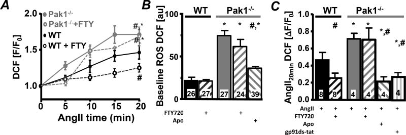
Fig. 3. Attenuated Pak1 activity increases ROS.
(A) Change in fluorescence recorded in WT (black) and Pak1−/− (grey) AMs loaded with DCF during stimulation with AngII in the absence or presence of FTY720 (open symbols). Bar graphs show DCF fluorescence at baseline (B) and (C) 20 min after AngII, AngII+FTY720, AngII+Apo (Apo: 1 µmol/L) or AngII+gp91ds-tat (1 µmol/L) superfusion in WT and Pak1−/− cells. (*: p < 0.05 compared to WT; #:p < 0.05 compared to Ctrl).

