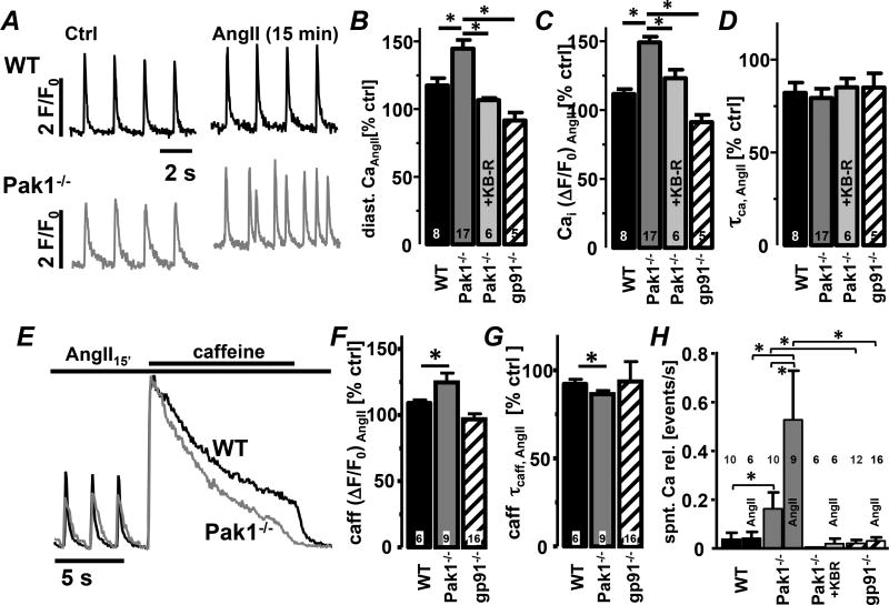
Fig. 6. Pak1−/− AMs exhibit exaggerated AngII-induced spontaneous Ca2+ transients.
(A) Field stimulation-induced Ca2+ transients from WT and Pak1−/− (grey) AMs. Bar graphs represent percent change after 15 min of AngII superfusion of (B) diastolic Ca2+, (C) Ca2+ transient amplitude and (D) decay constant (τcaff) in AMs from WT, Pak1−/−, gp91phox−/−, and Pak1−/− AMs treated with KB-R7943 (KBR: 1 µmol/L). (E) Normalized caffeine transients (10 mmol/L) after 15 min of AngII superfusion. Bar graphs show (F) caffeine transient amplitude and (G) decay constant. (H) Mean number of spontaneous Ca2+ transients (events/s) in AMs before and after AngII superfusion for treatment conditions described in A–D. (*: p < 0.05)

